
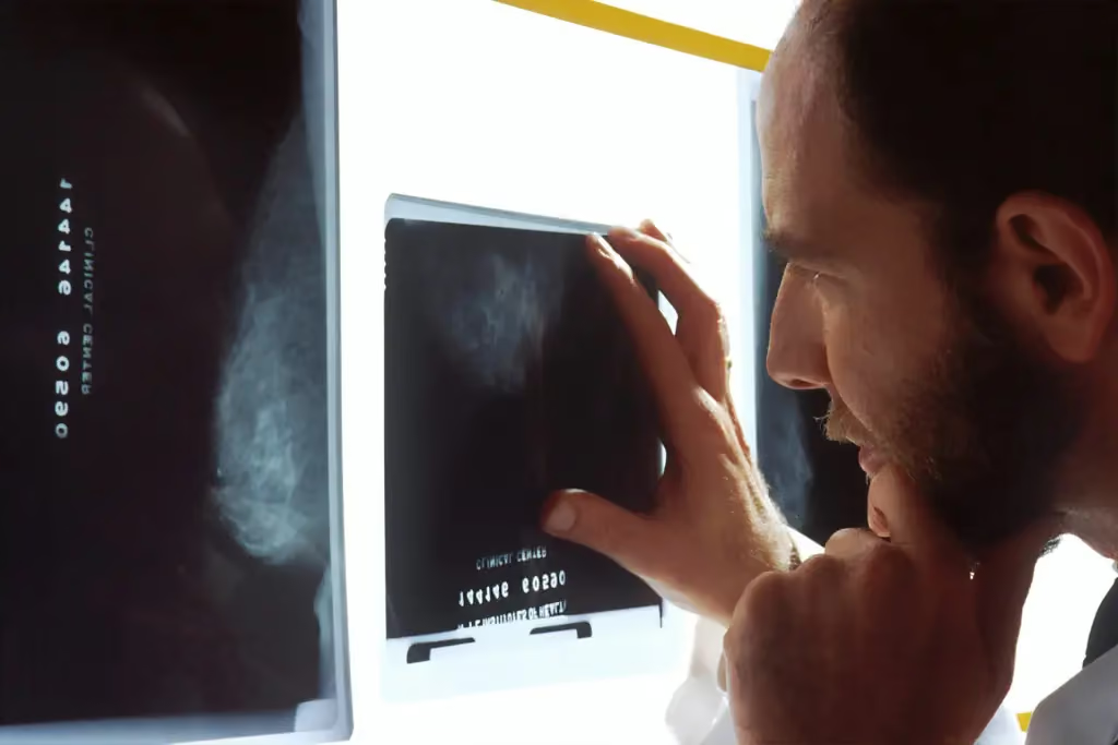
When it comes to diagnosing health issues in our beloved pets, MRI images have revolutionized the landscape of veterinary medicine. Did you know that these sophisticated imaging techniques can capture intricate details of soft tissues, providing invaluable insights into conditions that might otherwise go unnoticed? As veterinary practices increasingly embrace the use of MRI, the ability to accurately assess the health of animals before anesthesia or surgery has never been more critical.
This blog post will explore the significance of MRI images in veterinary anesthesia, delve into their benefits, limitations, and the future of this essential tool in enhancing animal health care. Whether you are a veterinarian looking to expand your knowledge or a pet owner interested in the intricacies of medical imaging, understanding the role of MRI images is imperative for effective and safe veterinary practices.
What Are MRI Images in Veterinary Medicine?
Magnetic Resonance Imaging (MRI) is a powerful diagnostic tool used in veterinary medicine, offering a non-invasive way to visualize the internal structures of animals. MRI images are created using strong magnets and radio waves, providing detailed images of soft tissues, organs, and the central nervous system. This imaging technique has revolutionized the ability to identify medical conditions in pets.
Importance of MRI Images for Pet Health
MRI images play a crucial role in diagnosing various health issues in animals. They are particularly useful for conditions that involve soft tissues, which may not be easily seen on X-rays or ultrasound. Here are some key benefits:
- Detailed Visualization: MRI provides high-resolution images, allowing veterinarians to identify tumors, spinal cord injuries, or brain abnormalities.
- Non-invasive Approach: Unlike some procedures, MRIs do not require surgical intervention, reducing the risk for the animal.
- Comprehensive Diagnosis: MRI images help veterinarians detect and monitor the progression of diseases, enabling more effective treatment plans.
Understanding these images can lead to better decision-making, especially when planning veterinary anesthesia, where precise knowledge of an animal’s health condition is vital.
The Role of MRI Images in Veterinary Anesthesia
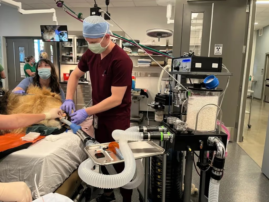
When it comes to veterinary anesthesia, accurate and comprehensive information about the animal’s health status is paramount. MRI images can significantly enhance this understanding, ultimately contributing to safer anesthesia practices.
How MRI Images Assist in Diagnosing Conditions
The diagnostic capabilities of MRI images allow veterinarians to:
- Identify underlying conditions that may complicate anesthesia, such as heart disease or neurological disorders.
- Assess the severity of conditions like tumors, leading to tailored anesthetic approaches.
- Determine the exact location and extent of injuries, which is crucial for surgical interventions.
By incorporating MRI images into the planning stages, veterinarians can make informed decisions, ensuring the safest possible environment for animals undergoing anesthesia.
The Impact of MRI on Anesthesia Planning
Effective anesthesia planning relies heavily on precise diagnosis. MRI images provide vital information that can influence:
- Anesthetic Drug Selection: Knowing the specific health issues can guide veterinarians in choosing appropriate anesthetics that minimize risk.
- Monitoring Requirements: MRI findings can dictate the level of monitoring needed during anesthesia, adding a layer of safety.
- Recovery Protocols: Understanding an animal’s unique medical condition allows for tailored post-anesthesia recovery plans.
In summary, MRI images are essential in veterinary practices for diagnosing health issues and enabling safer and more effective anesthesia management. By leveraging these advanced imaging techniques, veterinarians can provide the highest quality of care for their patients.
Understanding MRI Images: Key Terminology
To effectively utilize MRI images in veterinary medicine, it’s essential to understand the terminology associated with this imaging technique. Familiarity with key terms not only helps veterinarians but also educates pet owners about the importance of these images in diagnosing and managing health issues.
Common Terms Associated with MRI Images
- T1 and T2 Weighted Images: These terms refer to different types of MRI images that highlight various tissue properties. T1-weighted images provide contrast for fat-rich tissues, while T2-weighted images are better for showcasing fluids, such as edema or cysts.
- Slice: This term refers to a single image layer obtained during an MRI scan. Multiple slices are combined to create a three-dimensional representation of the area being examined.
- Contrast Agents: These are substances used to enhance the visibility of certain tissues during MRI imaging. They are particularly helpful in identifying tumors or inflammatory conditions.
- Artifacts: These are discrepancies in MRI images caused by various factors, such as motion or equipment malfunction. Understanding artifacts is crucial for accurate interpretation of scanned images.
Types of Tissues Visible in MRI Scans
MRI images can distinctly visualize several types of tissues, making it an invaluable diagnostic tool in veterinary medicine:
- Soft Tissues: MRI excels at imaging soft tissues, including muscles, fat, and connective tissues, which are often difficult to evaluate using other imaging techniques.
- Central Nervous System: Brain and spinal cord structures can be distinctly visualized, allowing for the diagnosis of conditions like tumors or degenerative diseases.
- Joint Structures: MRI can reveal abnormalities in ligaments and cartilage, helping to diagnose conditions such as cruciate ligament tears or osteoarthritis.
By understanding these terms and the types of tissues visible in MRI images, both veterinarians and pet owners can appreciate the significant role these images play in diagnosing animal health issues.
For more detailed explanations and resources related to MRI technology, you can visit Mallard Medical for the latest advancements and applications in the field.
Interpreting MRI Images: A Step-by-Step Approach
Interpreting MRI images can appear daunting due to their complexity. However, a systematic approach can simplify the process and enhance the accuracy of the diagnosis. Understanding how to read these images is crucial for veterinarians in determining the best treatment options for their patients.
Basic Anatomy Visible in MRI Images
MRI images provide a wealth of information about the anatomy of the animal. Familiarizing yourself with the basic structures can help in identifying abnormalities:
- Brain Structures: The brain’s various regions, including the cerebrum, cerebellum, and brainstem, are clearly delineated in MRI images.
- Spinal Cord: The entire vertebral column and spinal cord are visible, making it easier to identify conditions such as disc herniation or spinal tumors.
- Muscles and Tendons: MRI effectively shows muscle groups and tendons, allowing veterinarians to assess injuries or conditions like tendonitis.
Common Abnormalities and What They Indicate
Recognizing common abnormalities in MRI images can lead to earlier and more accurate diagnoses. Here are a few key issues veterinarians often encounter:
- Tumors: Abnormal growths can appear as irregular masses, highlighting the need for further investigation.
- Fluid Accumulation: Areas with increased fluid may indicate inflammation, infection, or cyst formation.
- Degenerative Changes: MRI can reveal signs of degeneration in joints, such as cartilage loss or bone spurs, crucial for conditions like osteoarthritis.
A Diagnostic Framework
To effectively interpret MRI images, consider using the following framework:
- Identify the Region: Determine the area of interest based on clinical signs and history.
- Examine the Basic Anatomy: Familiarize yourself with normal structures before assessing for abnormalities.
- Look for Pathology: Focus on unusual shapes, sizes, and textures within the images.
- Correlate with Clinical Findings: Integrate the imaging results with physical examination findings and lab tests for a comprehensive assessment.
Through this step-by-step approach, veterinarians can enhance their ability to interpret MRI images, facilitating accurate diagnoses and better treatment planning.
Benefits of Using MRI Images in Veterinary Anesthesia
The integration of MRI images in veterinary anesthesia offers numerous advantages that contribute to both the safety and effectiveness of surgical procedures. Understanding these benefits can help veterinarians make informed decisions when planning anesthesia for their patients.
Improved Accuracy in Diagnosis
One of the primary benefits of utilizing MRI images is their clarity and detail, which significantly enhances diagnostic accuracy. Key points include:
- Detailed Soft Tissue Visualization: MRI excels in imaging soft tissues that are often difficult to assess with other imaging techniques, allowing for the early detection of conditions such as tumors or infections.
- Comprehensive Evaluation: MRI images provide a thorough understanding of anatomical structures, aiding in the diagnosis of complex cases and reducing the likelihood of misdiagnosis.
Enhanced Safety During Anesthesia Procedures
MRI imaging also contributes to increased safety during anesthesia. This is particularly important in high-risk cases. Consider the following safety implications:
- Informed Anesthetic Protocols: With precise information from MRI images, veterinarians can tailor their anesthetic choices and dosages based on the specific needs of the patient.
- Improved Monitoring: Understanding the underlying health conditions prior to anesthesia allows for enhanced monitoring protocols, ensuring that veterinary teams can respond more effectively to any complications that may arise.
- Prevention of Complications: Detailed imaging can reveal pre-existing conditions that may complicate anesthesia, allowing veterinarians to mitigate risks before surgical procedures.
Better Surgical Outcomes
Ultimately, the use of MRI images can improve surgical outcomes in veterinary practices. Some benefits include:
- Effective Surgical Planning: MRI images help veterinarians strategize surgical approaches, especially in complicated cases that involve multiple structures.
- Reduced Surgery Time: Increased knowledge through imaging can streamline procedures, reducing time under anesthesia and enhancing recovery.
Incorporating MRI images into the anesthesia planning process not only facilitates safer procedures but also improves overall patient care.
Limitations of MRI Images in Veterinary Anesthesia
While MRI images are invaluable in veterinary medicine, they are not without limitations. Understanding these constraints is essential for veterinarians to make well-informed decisions regarding diagnostics and treatment plans.
Situations Where MRI May Not Be Effective
Though MRI images provide detailed insights, there are specific scenarios where they may fall short:
- Cost and Accessibility: MRI machines are expensive to operate and maintain, which can limit access in some veterinary practices, particularly in rural areas. This accessibility issue may delay diagnosis and treatment for pets in need.
- Time Consumption: Acquiring MRI images can take longer than other imaging modalities, such as X-rays or ultrasounds. This delay may not be suitable for emergency situations where immediate diagnostics are required.
- Motion Artifacts: Animals, especially those that are anxious or in pain, may have difficulty remaining still during the imaging process. This can result in motion artifacts that obscure important details in the MRI images, complicating interpretation.
Challenges in Accessing MRI Technology
In addition to the limitations of MRI itself, there are other barriers that can impact its use in veterinary anesthesia:
- Equipment Limitations: Not all veterinary facilities are equipped with MRI machines, meaning some veterinarians may need to refer cases to specialized centers, which can slow down the diagnosis process.
- Anesthesia Requirements: In some cases, animals may need anesthesia to remain still for the MRI scan, creating potential risks in cases with comorbid health conditions.
Understanding the Balance
Despite these limitations, MRI images remain a powerful diagnostic tool in veterinary medicine. Veterinarians are encouraged to balance the advantages of MRI imaging with its limitations to make the best possible decisions for their patients.
By staying informed about the potential hurdles associated with MRI, veterinarians can better evaluate when to utilize this advanced imaging technique.
The Future of MRI Imaging in Veterinary Medicine
As technology advances, the field of veterinary medicine continues to evolve, particularly regarding imaging techniques such as MRI. The future of MRI imaging holds promising developments that will enhance its utility in veterinary anesthesia and overall animal healthcare.
Advancements in MRI Technology
Recent innovations in MRI technology are set to improve both the quality of images and the speed of diagnostics. Key advancements include:
- Higher Resolution Imaging: New MRI machines are capable of producing finer images with greater clarity, allowing for better visualization of small structures and abnormalities in soft tissues.
- Faster Scanning Times: Innovations in MRI protocols are leading to reduced scanning times, which can decrease the need for anesthesia and make the process less stressful for animals.
- Functional MRI: This emerging technology allows for the assessment of brain activity by detecting changes in blood flow. It holds potential for evaluating neurological conditions in pets, further bridging the gap between medical imaging and functional analysis.
Potential Impact on Veterinary Anesthesia Practices
The advancements in MRI technology can significantly influence veterinary anesthesia practices in the following ways:
- Enhanced Preoperative Planning: Improved imaging techniques will help veterinarians make more informed decisions regarding anesthetic protocols, contributing to safer surgical outcomes.
- Early Detection of Health Issues: With better imaging, conditions may be detected earlier, allowing for timely interventions and tailored anesthesia plans that accommodate the animal’s specific needs.
- Increased Access to MRI Services: As technology becomes more cost-effective, more veterinary practices may incorporate MRI imaging, making it more accessible to a broader range of pet owners.
The Role of Education and Training
As MRI technology continues to advance, education and training for veterinarians will become increasingly important. Understanding how to effectively utilize and interpret the latest MRI images will be crucial for maximizing benefits in clinical practice.
- Continued Professional Development: Veterinarians should engage in ongoing education to stay updated on the latest advancements in MRI technology and imaging techniques.
- Collaboration with Radiologists: Building relationships with veterinary radiologists can provide veterinarians with additional support in interpreting complex MRI images effectively.
The future of MRI imaging in veterinary medicine is bright, with the potential to improve diagnostics, enhance safety in anesthesia, and ultimately provide better care for our animal companions.
Conclusion
In summary, MRI images have become a cornerstone in veterinary medicine, significantly enhancing diagnostics, especially in the context of anesthesia. Understanding the intricacies of MRI technology, its advantages, and its limitations is crucial for veterinarians aiming to provide optimal care to their patients.
Key Takeaways
- Enhanced Diagnostic Capabilities: MRI images allow for detailed visualization of soft tissues, aiding in the early detection of various health issues and improving overall treatment outcomes.
- Safe Anesthesia Practices: The accurate insights provided by MRI imaging help veterinarians tailor their anesthetic approaches, ensuring a safer environment during surgical procedures.
- Ongoing Advances: The future of MRI technology promises further improvements in imaging quality, speed, and accessibility, offering exciting possibilities for veterinary practices.
Emphasizing Education and Collaboration
To fully leverage the benefits of MRI images, continuous education for veterinarians is essential. Collaborating with radiology specialists can provide critical insights that enhance the interpretation of these images, allowing for more comprehensive care.
As the veterinary field continues to evolve, embracing advanced imaging modalities like MRI will become increasingly important. These technologies not only enrich the diagnostic toolkit for veterinarians but also ensure better health outcomes for our beloved animal companions.
The Importance of Combining MRI Images with Clinical Assessments
While MRI images are a powerful tool in diagnosing and managing veterinary conditions, they work best when combined with thorough clinical assessments. This integration enhances the overall treatment approach and ensures that veterinarians can provide comprehensive care for their patients.
The Role of Clinical Assessments
Clinical assessments provide the foundational context necessary for interpreting MRI images accurately. Key components include:
- Clinical History: Understanding the patient’s medical background and presenting symptoms is crucial. This information guides the interpretation of the MRI images and helps in identifying potential conditions.
- Physical Examination: A thorough physical examination can reveal aspects of an animal’s health that may not be visible in MRI images alone. It helps to confirm or rule out conditions hinted at by imaging.
- Laboratory Tests: Blood work and other diagnostic tests provide valuable data that can correlate with findings from MRI images, allowing for a more comprehensive understanding of the animal’s health.
Enhancing Diagnostic Accuracy
Combining MRI images with clinical assessments results in several significant benefits:
- Improved Diagnostic Confidence: When clinical findings align with MRI results, veterinarians can be more confident in their diagnoses, leading to more effective treatment plans.
- Holistic Patient Evaluation: A comprehensive approach ensures that all aspects of a pet’s health are considered, leading to better management of complex cases where multiple issues may exist.
- Tailored Treatment Plans: By integrating both imaging and clinical data, veterinarians can develop personalized treatment plans that address the individual needs of the animal.
Collaborative Approach in Veterinary Medicine
Encouraging collaboration among veterinary professionals can enhance the utilization of MRI images in practice. Strategies to promote teamwork include:
- Interdisciplinary Discussions: Regular case discussions involving veterinarians, radiologists, and other specialists can foster a richer understanding of patient cases.
- Continuous Education: Workshops and training sessions focused on combining imaging techniques with clinical methods can improve diagnostic skills and treatment outcomes.
In conclusion, the synergy between MRI images and clinical assessments is essential in veterinary medicine. By leveraging both aspects, veterinarians can ensure a more accurate and effective approach to diagnosing and treating conditions in animals.
Frequently Asked Questions
What are MRI images, and how are they used in veterinary medicine?
MRI images are non-invasive diagnostic tools that use strong magnetic fields and radio waves to create detailed images of soft tissues in animals. In veterinary medicine, they are used to assess organs, muscles, and the nervous system, providing crucial information for diagnosing health issues that are not easily visible through other imaging techniques.
Are MRI scans safe for pets?
Yes, MRI scans are generally safe for pets. They do not use ionizing radiation, which is a concern with some other imaging modalities like X-rays. However, pets may require sedation to keep them still during the procedure, which comes with its own set of risks that veterinarians carefully evaluate.
How long does an MRI scan take?
The duration of an MRI scan can vary depending on the area being examined and the specific protocol used, but it typically takes between 30 minutes to an hour. This includes preparation time as well as the actual imaging process. More complex cases may require additional time.
What are the limitations of MRI images in veterinary anesthesia?
While MRI images provide detailed insights, they do have limitations. These include potential motion artifacts caused by animals not remaining still during the scan, accessibility issues due to the high cost of MRI machines, and the necessity for sedation, which can pose risks to certain patients.
How can veterinarians improve their use of MRI images in practice?
Veterinarians can enhance their use of MRI images by undergoing continual education and training in imaging techniques, collaborating with radiology specialists for complex cases, and integrating MRI findings with clinical assessments for a more comprehensive understanding of their patients’ health.